Digital Dental Imaging Centers in Chelsea: Advanced Diagnostics for Complex Cases
Digital dental imaging centers in Chelsea represent a significant advancement in diagnostic capabilities for complex cases. By employing technologies like Cone Beam Computed Tomography (CBCT) and digital radiography, these centers provide high-resolution images essential for precise treatment planning. The integration of sophisticated imaging software allows for detailed anatomical analysis, promoting effective collaboration among multidisciplinary teams. Such innovations not only optimize patient outcomes but also redefine dental care efficiency, particularly in Chelsea’s urban landscape. What further implications might this evolution hold?
The Evolution of Dental Imaging Technology
As dental imaging technology has advanced, the field has undergone significant transformations, revolutionizing diagnostic capabilities and patient care. Historically, the shift from traditional film-based radiography to digital systems marked a pivotal moment. This shift enabled enhanced image quality, reduced radiation exposure, and expedited processing times. The integration of Computer-Aided Design (CAD) and Computer-Aided Manufacturing (CAM) systems further exemplified the technological integration within dentistry, facilitating precise treatment planning and execution. Additionally, the incorporation of Cone Beam Computed Tomography (CBCT) provided three-dimensional imaging, offering unparalleled insights into complex anatomical structures. These historical advancements underscore the progressive nature of dental imaging technology. As a result, practitioners now possess enhanced diagnostic tools, contributing to improved patient outcomes and streamlined clinical workflows in modern dental practices.
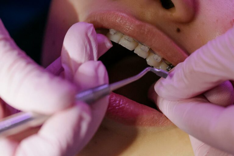
Understanding Digital Radiography and Its Benefits
Digital radiography in dental practice offers significant advancements in image clarity and diagnostic precision, attributed to its superior resolution and contrast capabilities. This technology also results in a substantial reduction in radiation exposure, enhancing patient safety without compromising diagnostic efficacy. As a result, digital radiography presents a compelling case for widespread adoption in modern dental imaging centers.
Enhanced Image Quality
When considering advancements in dental technology, digital radiography stands at the forefront due to its ability to produce enhanced image quality. This technology notably improves image resolution, offering dentists a precise view of dental structures. Enhanced image resolution translates into visual clarity that is paramount for accurate diagnosis and treatment planning. Digital sensors capture images with greater detail compared to traditional film-based methods, allowing for the identification of minute anomalies that may not be visible otherwise. The ability to manipulate image parameters, such as contrast and brightness, further enhances visual clarity, facilitating an extensive analysis. Consequently, digital radiography provides practitioners with a superior diagnostic tool, improving the efficacy of dental assessments in complex cases. This advancement underscores the importance of investing in cutting-edge imaging technology.
Reduced Radiation Exposure
While traditional radiographic methods have served dentistry for decades, digital radiography offers a distinct advantage by considerably reducing patient radiation exposure. This advancement prioritizes radiation safety, a paramount concern within dental diagnostics. By employing digital sensors, the radiation dose is minimized, thereby enhancing patient comfort and alleviating potential apprehensions associated with conventional X-ray procedures. The utilization of digital radiography in Chelsea’s dental imaging centers underscores a commitment to reducing overall radiation risks while maintaining diagnostic efficacy. Moreover, the immediate processing capability of digital systems reduces the need for retakes, additionally diminishing radiation exposure. This technological evolution represents a significant step forward in dental diagnostics, aligning with modern healthcare’s emphasis on safety and patient-centric care. Such advancements guarantee ideal diagnostic outcomes with minimal radiation impact.
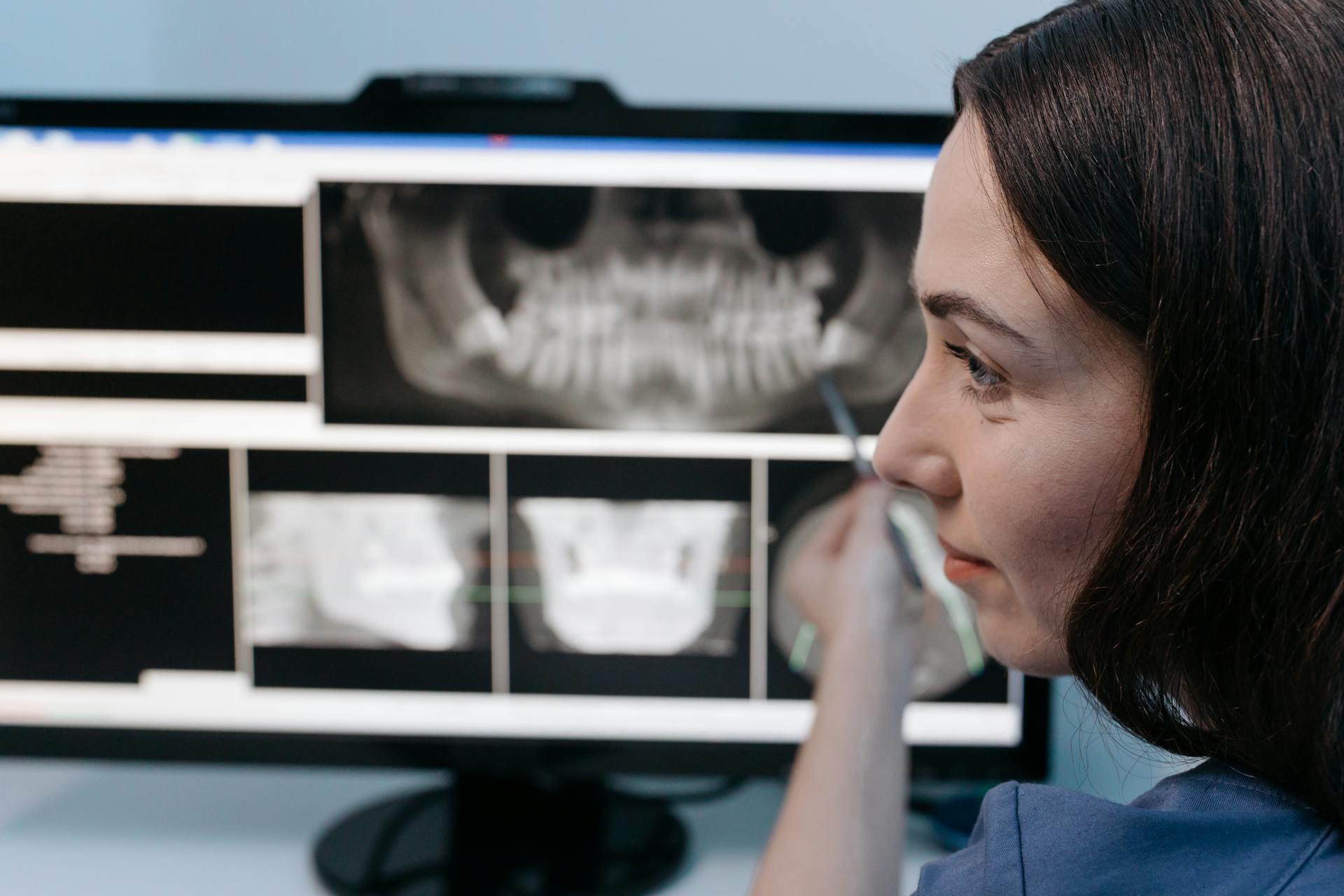
Exploring 3D Imaging Techniques in Dentistry
Three-dimensional imaging techniques have revolutionized modern dentistry, offering profound enhancements in diagnostic accuracy and treatment planning. Utilizing 3D scanning, these techniques provide an extensive view of dental structures, facilitating the detailed visualization of complex anatomical features. This approach enables practitioners to conduct volumetric analysis, which is essential for evaluating the spatial relationships and dimensions within the oral cavity. It allows for precise measurements that are not achievable with traditional two-dimensional imaging methods. By integrating advanced software, dental professionals can manipulate and examine these images from multiple angles, ensuring a more thorough examination of potential issues. As a result, 3D imaging plays a pivotal role in the development of tailored treatment plans, ultimately improving patient outcomes and enhancing the overall quality of care.
How Digital Imaging Enhances Diagnostic Accuracy
How precisely does digital imaging enhance diagnostic accuracy in dentistry? The integration of advanced imaging software facilitates the generation of highly detailed, three-dimensional models of dental structures. These models allow practitioners to detect anomalies with increased precision. AI integration plays a pivotal role by analyzing vast datasets to identify subtleties that may escape human observation, such as minute fractures or incipient carious lesions. This technology reduces diagnostic errors, ensuring more reliable outcomes. Additionally, digital imaging provides instantaneous results, accelerating the diagnostic process. Enhanced image clarity and resolution surpass the capabilities of traditional radiography, offering superior detail. As a result, dental professionals can make informed decisions based on thorough data analysis, thereby improving the accuracy of diagnoses and contributing to more effective patient care strategies.
Tailoring Treatment Plans With Advanced Imaging
Leveraging advanced imaging technologies considerably optimizes the development of personalized treatment plans in dentistry. Imaging precision allows practitioners to capture detailed anatomical information, facilitating an extensive understanding of each patient’s unique dental structure. This high-resolution data is critical for treatment customization, enabling clinicians to tailor interventions specifically suited to individual needs. Digital imaging modalities, such as CBCT (Cone Beam Computed Tomography), offer volumetric insights, revealing intricate details that traditional methods may overlook. Consequently, practitioners can identify anatomical variations, pathologies, and spatial relationships with unprecedented accuracy. This precision guarantees that treatment strategies are not only effective but also minimally invasive, enhancing patient outcomes. The application of advanced imaging fosters a more targeted approach, reducing unnecessary interventions and consequently optimizing overall care delivery.
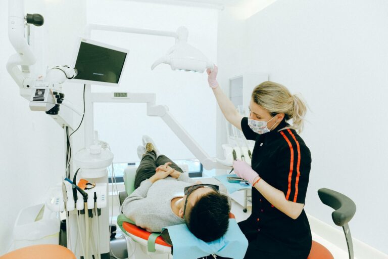
The Role of Imaging Centers in Complex Dental Cases
In the domain of complex dental cases, imaging centers play a pivotal role by providing indispensable diagnostic capabilities that markedly enhance clinical decision-making. These centers facilitate a collaborative diagnosis by offering advanced imaging technologies such as cone beam computed tomography (CBCT) and digital radiography. Such modalities deliver high-resolution images that are critical for accurate evaluation of intricate dental structures. Through precise imaging, practitioners can engage in effective case management, addressing conditions like impacted teeth, jaw deformities, and complex endodontic issues with greater accuracy. Moreover, imaging centers serve as a nexus for multidisciplinary teams, enabling dentists, oral surgeons, and orthodontists to synchronize their efforts. This integration guarantees each expert contributes to a thorough treatment plan, optimizing patient outcomes in challenging dental scenarios.
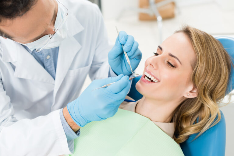
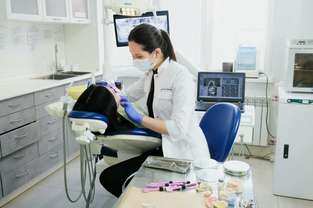
Patient Experience in Digital Dental Imaging Centers
Within the landscape of digital dental imaging centers, the patient experience is meticulously crafted to guarantee efficiency and comfort, underscoring the facility’s commitment to high-quality care. Central to this approach is the integration of state-of-the-art technology that enhances patient comfort through reduced imaging times and minimal radiation exposure. Imaging accessibility is prioritized by facilitating easy appointment scheduling and expeditious image processing. The centers employ ergonomic equipment designed to accommodate diverse patient needs, further contributing to a seamless experience. Additionally, staff are trained to provide clear communication and support, ensuring patients understand procedures and outcomes. This strategic focus on patient-centric care elevates the overall experience, aligning with the centers’ objective of delivering precise diagnostics in a patient-friendly environment.
The Impact on Dental Professionals and Their Practice
Digital dental imaging centers greatly influence dental professionals and their practices by revolutionizing diagnostic processes and enhancing treatment precision. These centers provide advanced tools that streamline practice management through efficient workflow integration, reducing diagnostic time and improving patient turnover. By utilizing sophisticated imaging technologies, professionals can deliver more accurate diagnoses, leading to better treatment outcomes and higher patient satisfaction.
Moreover, digital imaging fosters professional development by enabling practitioners to stay abreast of cutting-edge advancements, thereby enhancing their expertise. Access to high-quality images allows for improved case documentation and facilitates peer collaboration, furthering continuous learning and refinement of skills. Consequently, practitioners not only enhance their clinical acumen but also optimize practice operations, positioning themselves as leaders in quality care within the dental community.
The Future of Dental Imaging in Urban Settings
The future of dental imaging in urban settings is poised for significant advancements through the integration of innovative imaging technologies. These advancements are expected to enhance diagnostic accuracy, allowing practitioners to identify dental issues with greater precision. In addition, the implementation of these technologies is anticipated to increase urban dental efficiency by streamlining workflows and reducing patient wait times.
Innovative Imaging Technologies
As urban environments continue to evolve, the integration of innovative imaging technologies in dental practices is reshaping the landscape of patient care. Central to this transformation is the development of advanced imaging software, enabling precise diagnostics and treatment planning. Such software facilitates seamless imaging integration, allowing dental professionals to amalgamate various data sources into a coherent, thorough analysis. This integration leads to enhanced diagnostic accuracy, particularly in complex cases, by providing high-resolution, multi-dimensional visualizations of dental structures. In addition, these technologies support minimally invasive procedures by offering detailed insights that guide surgical interventions. As a result, patient outcomes improve due to more accurate diagnostics and targeted treatment strategies. Consequently, innovative imaging technologies are pivotal in advancing urban dental care efficiency and effectiveness.
Urban Dental Efficiency
Building upon the advancements in imaging technologies, urban dental practices are poised to achieve unprecedented levels of efficiency. The integration of digital imaging systems facilitates streamlined workflows, reducing patient wait times and enhancing throughput. Urban accessibility is greatly enhanced as these practices can serve a larger demographic, optimizing space and resource utilization. This is particularly crucial in dense metropolitan areas where patient volume is high. Additionally, digital imaging offers patient convenience through reduced appointment durations and immediate image availability. The capability for real-time data sharing allows for efficient interprofessional collaboration, further augmenting service delivery. As a result, urban dental centers can maintain high standards of care while accommodating the growing demand in city environments, marking a pivotal shift in dental service provision.
Enhanced Diagnostic Accuracy
With the advent of cutting-edge digital imaging technologies, urban dental practices are witnessing a significant enhancement in diagnostic accuracy. This progression is primarily driven by AI advancements and sophisticated imaging software that allow for more precise analysis of dental structures. These technologies facilitate early detection of pathologies, providing a higher resolution of images that surpasses conventional radiography. AI-powered algorithms enhance image interpretation, reducing human error and improving diagnostic confidence. By employing machine learning, these systems can detect subtle changes in dental anatomy that might be overlooked by traditional methods. Consequently, practitioners in urban settings are better equipped to manage complex cases, optimizing patient outcomes. The integration of these technologies represents a pivotal shift towards more accurate and efficient dental diagnostics.
Frequently Asked Questions
What Are the Costs Associated With Digital Dental Imaging Services in Chelsea?
The cost factors for digital dental imaging services in Chelsea vary, influenced by procedure complexity, equipment used, and practitioner expertise. Insurance coverage plays an essential role in determining out-of-pocket expenses for patients, impacting overall affordability.
Are Digital Dental Imaging Centers Open on Weekends?
Weekend availability of digital dental imaging centers enhances patient convenience by accommodating diverse schedules. Operational hours vary; some centers offer weekend services, ensuring accessibility for patients unable to attend appointments during standard weekday hours.
How Do Patients Access Their Digital Imaging Records?
Patients access their digital records through secure online portals, ensuring patient privacy. These systems require unique identifiers for login, safeguarding sensitive information. Access protocols adhere strictly to data protection regulations, maintaining confidentiality and integrity of patient records.
What Safety Measures Are in Place for Radiation Exposure?
Facilities implement radiation safety protocols, adhering strictly to exposure limits set by regulatory bodies. Advanced imaging technology minimizes radiation doses while protective equipment, such as lead aprons, shields patients, ensuring compliance with established safety standards.
Can Digital Imaging Detect Conditions That Traditional X-Rays Cannot?
Digital imaging, particularly 3D imaging, offers diagnostic advantages over traditional x-rays by providing detailed visualization of complex dental structures, aiding in the detection of conditions such as periodontal disease, bone loss, or impacted teeth more accurately and efficiently.