Digital Dental Imaging Chelsea: Advanced 3D Technology for Precise Diagnosis
Digital dental imaging represents a revolutionary advancement in modern dentistry, providing unprecedented diagnostic precision through cutting-edge technology like CBCT cone beam scans, digital X-rays, and intraoral cameras. Unlike traditional film-based radiography, these advanced systems deliver immediate, high-resolution images with up to 90% less radiation exposure, enabling same-day diagnosis and treatment planning for complex dental conditions.
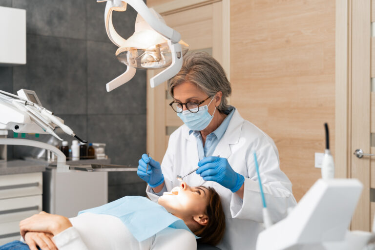
In Chelsea Manhattan, patients now have access to state-of-the-art imaging technology that transforms how dental professionals diagnose everything from hidden infections to complex anatomical variations. Whether you’re considering dental implants, need precise surgical planning, or require comprehensive TMJ analysis, digital imaging provides the detailed 3D visualization necessary for optimal treatment outcomes. At our Chelsea practice, we’ve invested in the most advanced imaging equipment to ensure our patients receive the highest standard of diagnostic care with maximum safety and comfort.
This comprehensive guide explores the various types of digital dental imaging available, when advanced 3D CBCT scans become necessary, and how these technologies benefit patients seeking superior dental care in the Chelsea area. Understanding these diagnostic tools empowers you to make informed decisions about your oral health while appreciating the precision and safety that modern dental imaging provides.
What Is Digital Dental Imaging Technology?
Digital dental imaging encompasses a suite of advanced technologies that capture, process, and display detailed images of your teeth, jawbone, and surrounding structures using electronic sensors and sophisticated computer systems. This revolutionary approach has largely replaced traditional film-based radiography, offering superior image quality, immediate results, and significantly reduced radiation exposure for enhanced patient safety.
The fundamental principle behind digital imaging involves converting X-ray energy into digital data through specialized sensors or detectors. This electronic capture eliminates the need for chemical film processing, allowing dental professionals to view images instantly on computer screens where they can be enhanced, magnified, and analyzed with unprecedented precision.
Types of Advanced Digital Imaging
Modern dental practices utilize several distinct digital imaging technologies, each designed for specific diagnostic purposes and clinical applications.
CBCT Cone Beam CT Scanning
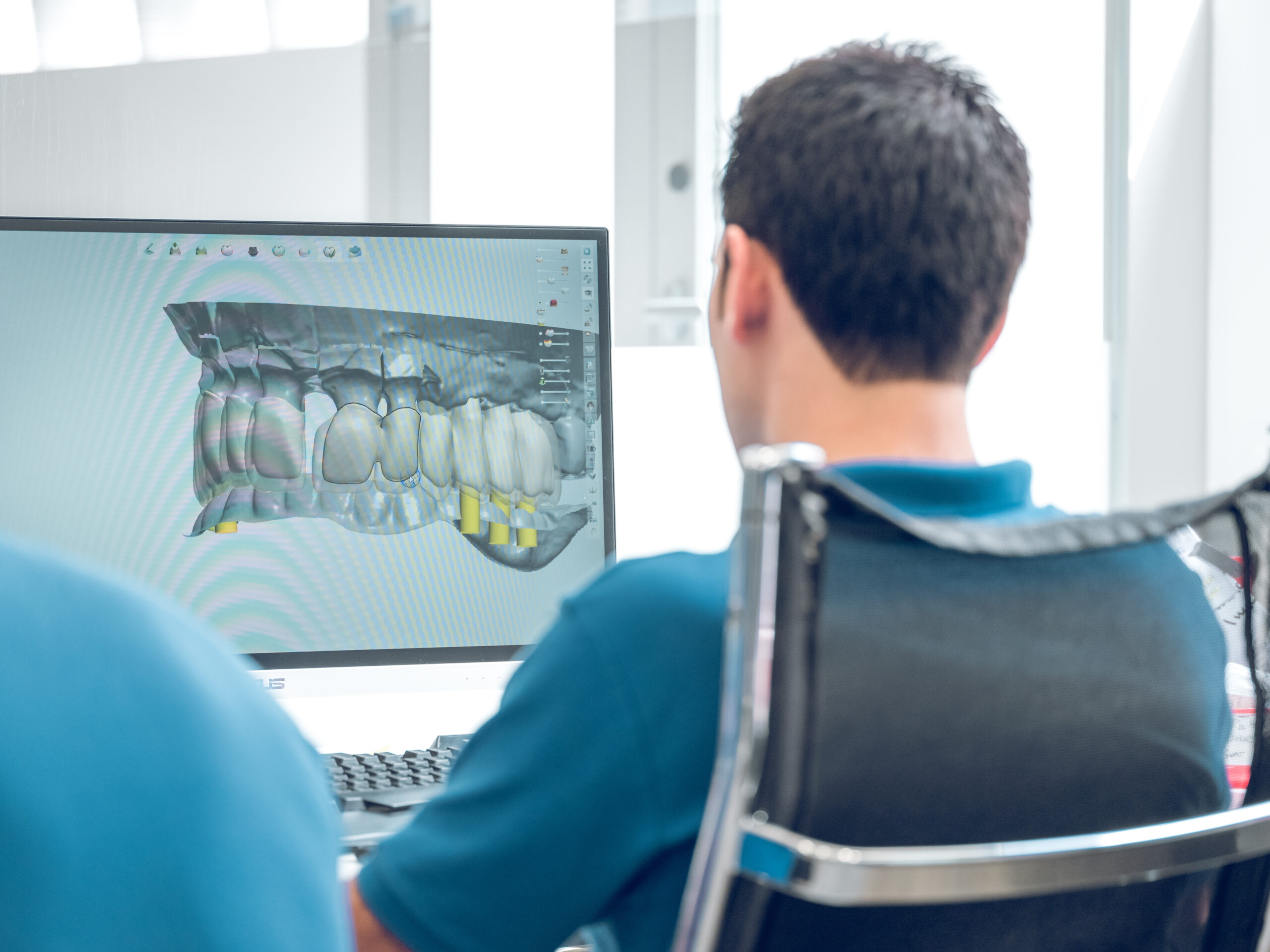
Cone Beam Computed Tomography (CBCT) represents the pinnacle of dental imaging technology, producing detailed three-dimensional images of your entire oral and maxillofacial region. This advanced system captures hundreds of individual images in a single rotation, which are then reconstructed by computer software into comprehensive 3D models showing bone density, nerve pathways, sinus cavities, and anatomical structures with remarkable clarity.
CBCT scanning proves invaluable for complex procedures requiring precise spatial relationships, such as dental implant placement, impacted tooth extraction, and TMJ analysis. The technology enables dentists to virtually plan treatments, create surgical guides, and identify potential complications before beginning actual procedures.
Digital X-Ray Systems
Digital X-ray technology utilizes electronic sensors to capture traditional two-dimensional radiographic images with exceptional speed and clarity. These systems produce images immediately after exposure, eliminating the wait time associated with film development while providing superior contrast and detail resolution.
The radiation reduction achieved through digital X-rays is particularly significant, with most systems delivering 80-90% less radiation exposure compared to traditional film radiography. This safety enhancement, combined with the ability to digitally enhance images for better visualization, makes digital X-rays the preferred standard for routine dental examinations.
Intraoral Camera Technology
Intraoral cameras represent a unique form of digital imaging that captures real-time, full-color images of your teeth and gums using a small, pen-like device inserted into your mouth. These cameras provide immediate visual documentation of dental conditions, enabling both dentist and patient to observe problems areas simultaneously on a computer monitor.
This technology excels at patient education, allowing you to see exactly what your dentist observes during examinations while creating detailed records for insurance documentation and treatment progress monitoring.
Revolutionary Benefits of Digital Dental Imaging
Digital imaging technology offers transformative advantages that benefit both patients and dental professionals through enhanced diagnostic capabilities, improved safety protocols, and streamlined treatment processes.
Immediate Diagnostic Results
The instant nature of digital imaging eliminates traditional waiting periods associated with film processing, enabling same-visit diagnosis and treatment planning. Your dentist can capture, review, and analyze images within minutes of exposure, allowing for immediate discussion of findings and treatment recommendations during your appointment.
This efficiency proves particularly valuable for emergency situations where rapid diagnosis determines treatment urgency. Additionally, immediate results prevent the need for return visits solely to review imaging findings, saving valuable time for busy Chelsea professionals and families.
Superior Image Quality and Precision
Digital sensors capture significantly more detail than traditional film, with the ability to electronically enhance images for optimal visualization. Dentists can adjust contrast, brightness, and magnification levels to examine specific areas more closely, potentially identifying problems that might be missed on conventional radiographs.
The precision afforded by digital imaging proves essential for complex procedures requiring exact measurements and spatial relationships. Whether planning dental implant placement or analyzing root canal anatomy, the enhanced detail and measurement capabilities of digital systems support more predictable treatment outcomes.
Radiation Safety and Dose Reduction
Perhaps the most significant advantage of digital imaging lies in its dramatic reduction of radiation exposure. Modern digital X-ray systems typically deliver 80-90% less radiation than traditional film radiography while maintaining superior image quality. This reduction addresses patient concerns about cumulative radiation exposure, particularly for individuals requiring frequent monitoring or multiple imaging sessions.
According to the American Dental Association, digital dental X-rays expose patients to radiation levels comparable to natural background radiation encountered in daily life, making them exceptionally safe for routine diagnostic use.
When Is Advanced 3D CBCT Imaging Necessary?
While digital X-rays suffice for routine dental examinations, certain complex conditions require the comprehensive visualization provided by CBCT cone beam scanning to ensure optimal treatment planning and execution.
Dental Implant Planning and Placement
CBCT imaging has become the gold standard for dental implant procedures, providing essential three-dimensional visualization of jawbone density, nerve pathways, and anatomical structures. This technology enables precise measurement of available bone volume, identification of critical anatomical landmarks like the inferior alveolar nerve, and virtual implant placement before actual surgery.
The 3D visualization capabilities allow dentists to create surgical guides that translate virtual planning into precise implant positioning, significantly improving success rates while minimizing surgical complications. At our Chelsea practice, we utilize CBCT technology for all implant cases to ensure optimal outcomes for our patients.
Complex Oral Surgery and Extractions
Impacted wisdom teeth, complex extractions, and oral surgical procedures benefit tremendously from CBCT imaging’s ability to reveal hidden anatomical variations and potential complications. The technology clearly shows root configuration, proximity to nerves and sinuses, and bone density variations that influence surgical approach and difficulty.
For patients requiring extraction of impacted teeth, CBCT scans provide crucial information about root proximity to adjacent teeth, nerve pathways, and sinus cavities, enabling surgeons to plan the safest and most efficient extraction technique.
TMJ Analysis and Airway Assessment
Temporomandibular joint (TMJ) disorders require detailed visualization of joint structures, bone relationships, and surrounding anatomy that only CBCT imaging can provide. This technology reveals joint space narrowing, bone changes, and anatomical variations that contribute to TMJ dysfunction.
Additionally, CBCT imaging supports airway analysis for patients with sleep apnea or breathing difficulties, showing the three-dimensional relationship between jawbone position and airway volume for treatment planning purposes.
Orthodontic Treatment Planning
Advanced orthodontic cases benefit from CBCT imaging’s ability to show root positions, bone levels, and anatomical variations that influence treatment planning. The technology proves particularly valuable for cases involving impacted teeth, root resorption concerns, or complex tooth movements requiring precise visualization of surrounding structures.
Is Advanced Digital Imaging Right for You?
Determining the appropriate level of imaging for your specific dental needs involves careful consideration of your symptoms, treatment requirements, and diagnostic challenges your dentist encounters during clinical examination.
Determining Your Imaging Needs
Your dentist considers multiple factors when recommending specific imaging modalities, including the complexity of your condition, treatment goals, and potential risks or complications. Routine dental care typically requires only digital X-rays, while complex restorative work, surgical procedures, or diagnostic challenges may necessitate advanced CBCT imaging.
The decision process involves weighing the diagnostic benefits against radiation exposure, cost considerations, and clinical necessity. Modern dental professionals follow evidence-based protocols to ensure imaging recommendations align with actual diagnostic requirements rather than routine protocols.
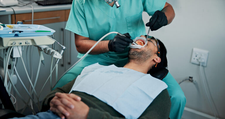
What to Expect During Digital Imaging
Digital X-ray procedures mirror traditional radiography but with enhanced comfort and speed. The electronic sensors used in digital systems are often more comfortable than traditional film holders, and the immediate results eliminate waiting periods associated with film processing.
CBCT scanning involves standing or sitting still while a rotating scanner captures images around your head, typically taking 20-40 seconds for complete image acquisition. The open design of most CBCT units minimizes claustrophobia concerns while providing comprehensive three-dimensional visualization of your oral structures.
Digital Imaging Safety and Radiation Protocols
Modern digital imaging prioritizes patient safety through advanced technology design and strict radiation protocols that minimize exposure while maximizing diagnostic value.
ALARA Radiation Safety Standards
Dental professionals follow ALARA (As Low As Reasonably Achievable) principles when utilizing any form of radiographic imaging. This approach ensures that radiation exposure remains at the minimum level necessary to achieve diagnostic objectives while maintaining image quality sufficient for accurate diagnosis and treatment planning.
Digital imaging technology inherently supports ALARA principles through its enhanced sensitivity and immediate feedback capabilities, which reduce the likelihood of repeat exposures due to technical factors or positioning errors.
Comparing Radiation Levels
To provide perspective on dental imaging radiation exposure, a typical digital dental X-ray delivers approximately 0.005 millisieverts (mSv) of radiation, comparable to the natural background radiation encountered during a few hours of daily life. Even CBCT scans, while delivering higher doses than traditional dental X-rays, provide radiation exposure similar to a few days of natural background radiation.
According to the FDA, these exposure levels remain well below thresholds associated with health concerns while providing invaluable diagnostic information for complex dental conditions.
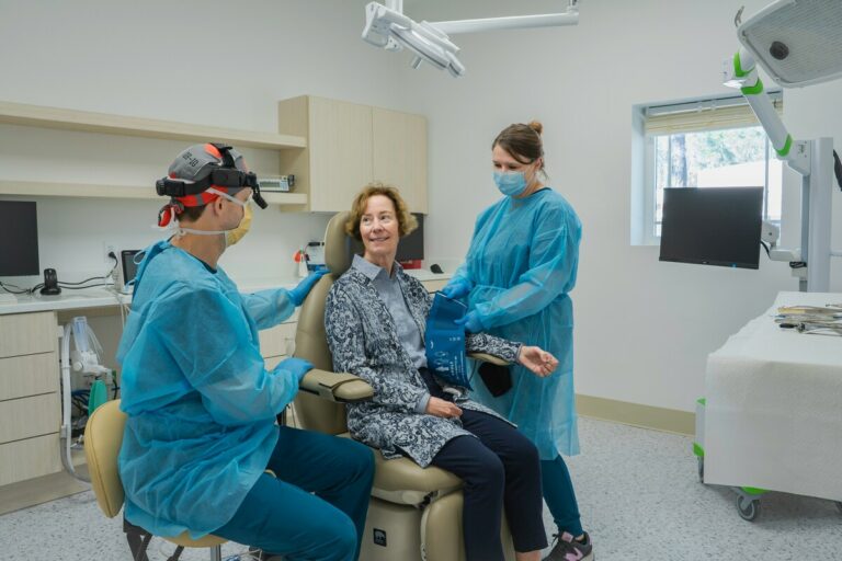
Advanced Digital Imaging in Chelsea Manhattan
Chelsea patients benefit from access to cutting-edge dental imaging technology through practices that have invested significantly in advanced diagnostic equipment and ongoing professional education to maximize imaging capabilities.
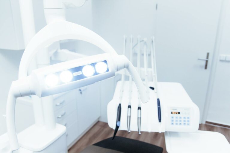
State-of-the-Art Equipment and Technology
Modern Chelsea dental practices utilize the latest generation imaging systems featuring enhanced resolution, reduced radiation exposure, and integrated software for comprehensive treatment planning. These technological investments reflect a commitment to providing patients with the highest standard of diagnostic care available in contemporary dentistry.
The integration of digital imaging with treatment planning software enables precise virtual procedures, surgical guide creation, and predictable treatment outcomes that benefit patients through reduced treatment time, improved comfort, and enhanced long-term success rates.
Convenient Chelsea Location and Same-Day Results
The central Manhattan location of Chelsea provides exceptional convenience for patients from surrounding areas including Flatiron, Midtown, and West Village, who can access advanced imaging technology without traveling to specialized imaging centers or university hospitals.
Same-day imaging results enable immediate treatment planning and decision-making, particularly valuable for busy professionals who prefer comprehensive dental care during single appointments rather than multiple visits for diagnosis and treatment planning phases.
Specialist Collaboration and Digital Records
Digital imaging facilitates seamless collaboration with dental specialists through immediate electronic image sharing and consultation capabilities. This integration ensures that complex cases receive appropriate specialist input while maintaining continuity of care within your primary dental practice.
The digital nature of these records also supports long-term monitoring of dental conditions, treatment progress tracking, and insurance documentation requirements while eliminating the storage and retrieval challenges associated with traditional film-based records.
Experience Advanced Digital Imaging Technology
Digital dental imaging represents a fundamental advancement in diagnostic capability that benefits Chelsea patients through enhanced precision, improved safety, and more efficient treatment planning. Whether you require routine dental care or complex restorative procedures, modern imaging technology provides the detailed visualization necessary for optimal outcomes with minimal radiation exposure and maximum convenience.
At our Chelsea practice, we’ve made significant investments in the most advanced digital imaging technology available to ensure our patients receive superior diagnostic care in a comfortable, convenient environment. Our experienced team utilizes these cutting-edge systems to provide same-day diagnosis, precise treatment planning, and seamless coordination with specialists when necessary. Contact us to schedule your consultation and experience the benefits of advanced digital imaging technology firsthand.